Contact us
- Macquarie Medical Imaging
- T: +61 (2) 9430 1100
- E: mmi.enquiries@mqhealth.org.au
Macquarie University Hospital and the Macquarie Centre
View mapsGet in touch or upload your existing referral
Learn moreAccess forms and fact sheets for referring patients to MMI
Referral informationPositron-Emission Tomography (PET) is a form of nuclear medicine imaging in which a small, safe amount of radioactive tracer – typically a simple form of glucose called fluorodeoxyglucose – is injected into the bloodstream.

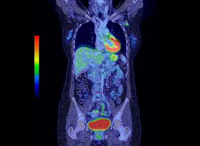
The PET scanner visualises the uptake of this tracer to create an image of the tissue and organs in the body. It can provide valuable information for the diagnosis and treatment of a variety of diseases like cancer, brain or heart disease. It’s also used to investigate epilepsy, Alzheimer’s disease, brain disorders, central nervous system problems, and heart disease.
PET scans are more sensitive in detecting cancer and other diseases than other imaging modalities. PET scanners have a built-in diagnostic CT so that the PET images can be fused (overlaid) perfectly one on the other. These fused images incorporate the sensitivity of a PET scan with the superior anatomy of a diagnostic CT.
By using a combined PET and CT scanner, MMI is able to provide high-resolution CT scans at the same time as a PET scan, saving you time and providing the most useful anatomical and functional information to guide treatment.
Watch video
The radioactive injection is given directly into your bloodstream via your vein. Different types of radioactive tracer target different types of disease. The PET scanner then detects the tracer and builds a digital image allowing your doctor see what’s going on in your tissues or organs.
The most commonly used form of radioactive tracer is radioactive glucose called FDG. Cancer cells absorb more glucose as compared to the normal cells around it to sustain its abnormal growth rate. The PET scan detects that there is more radiation coming from that area as opposed to the normal healthy cells.
Aside from being able to measure blood flow and oxygen intake, this procedure can check how your body uses sugar.
A PET scan is usually an outpatient procedure. It requires you to fast six hours before the scan.
In preparation for your scan, ensure that you:
Some additional tips to remember:
Once the radiotracer is injected, it usually takes up to one hour to course through your body and be absorbed into the organ or tissue to be checked. The scan takes around 30 minutes to an hour to complete.
The physician and radiologist then review the images and write a report, which is sent to your doctor in one to two days.
Whether you need a PET or a CT scan depends on your doctor’s recommendations. Here at Macquarie Medical Imaging, we do provide high-resolution CT scans at the same time as a PET scan, saving you time and providing the most useful anatomical and functional information to guide treatment.
Our PET/CT scanner is research-grade and operated by our experienced staff who are actively involved in research and clinical medical imaging.