Full-body 3D imaging with the lowest radiation dose
EOSedgetm allows for head to toe, frontal and lateral images to be acquired simultaneously, with the patient in either standing or seated position, without compromising image quality.
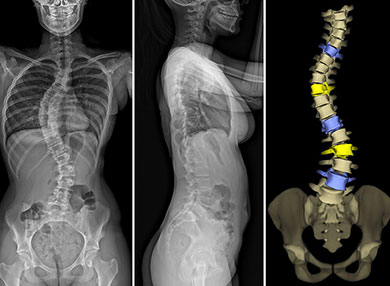
EOSedgetm is an innovative 2D / 3D imaging system for the diagnosis, treatment and monitoring of orthopaedic and osteoarticular pathologies, including those involving the hips, knees and spine. It is the first technology capable of providing full-body, 2D and 3D images of patients in a standing position at a low dose of radiation. It is suitable for both adults and children.
Reducing radiation dose is particularly beneficial for children requiring frequent imaging, such as children with spinal deformities like scoliosis.
All EOSedgetm scans at MMI are bulk-billed and no preparation is required.
EOSedgetm is a device that captures bi-planar images with two perpendicular X-rays beams that travel vertically while scanning the patient from head to toe.
In less than 20 seconds, the EOSedgetm exam produces simultaneous frontal and lateral, low dose images. The two resulting digital images are processed by the EOSedgetm proprietary sterEOS® software to generate a 3D model of the patient’s spine and/or lower limbs.
These 3D models provide highly detailed information about the patient’s unique anatomy to better assist orthopaedic and spinal surgeons as they diagnose patients.
This additional data can be used for precise 3D surgical planning to help improve overall patient outcomes by identifying patients at risk and developing a customised surgical plan.