What is nuclear medicine?
Nuclear medicine imaging uses a small amount of radioactive tracer to visualise how the body is functioning. It can assist doctors in the diagnosis or treatment of a variety of illnesses and conditions.
When it comes to nuclear medicine imaging, a specialist gives you a small amount of radioactive tracer. It’s either swallowed, inhaled, or injected into a vein or shunt, which then emits radioactive energy (gamma rays) for a short time.
The specialist then uses the gamma camera to detect the energy that’s emitted, and this creates images of how a certain body part or organ is functioning, giving doctors the precise information they need about the area being examined.
Common nuclear medicine procedures
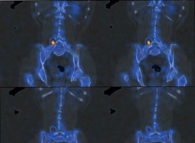
- Bone scan – This shows the effects of bone-related injury, disease, or infection. This also allows doctors to see how well the bone is functioning post-treatment
- Cardiac (sestamibi) – Cardiac nuclear medicine aids in diagnosing or assessing heart disease such as coronary artery disease, ischaemia or cardiomyopathy
- Gated heart pool – This involves identifying the red blood cells with a radiopharmaceutical and then measuring the amount of blood in it during different parts of the heartbeat.
- Liver scan – This helps doctors examine the liver and the spleen for potential diseases such as cancer, hepatitis, cirrhosis, etc.
- Biliary function – This helps assess biliary flow, cystic duct patency, and gallbladder function (ejection fraction)
- Gastric emptying – This measures your ability to digest food over time
- Reflux – When the doctor wants to look at excessive acid-containing secretions into the oesophagus
- Colon transit – This helps evaluate or diagnose constipation in parts of the large colon
- V/Q lung scan (with lung lobe separation and quantification) – This examines airflow and blood flow in the lungs and checks for any blood clots; it can also quantitate the function of each part of the lung.
- Thyroid scan – assess the thyroid gland; its activity, function, size, location and to evaluate thyroid nodes.
The gamma camera is a special camera that detects radioactive energy that your body gives off after taking in the radiotracer and records it as flashes of light. This is then converted into digital images of the part of the body under examination.
The gamma camera is highly sensitive to the physiological functioning of targeted cells in your body.
The gamma camera has an inbuilt CT that allows the sensitive images of the gamma camera to be fused (overlaid) perfectly with a high-quality CT. This fused image or SPECT/CT is the highest quality image as it incorporates the sensitivity of images generated by the gamma camera with the superior quality anatomy from a CT.
Preparing for a nuclear medicine exam varies. Prior to your scheduled exam, your attending physician will tell you what you need to do to prepare for your test.
If you are pregnant or breastfeeding you must inform your doctor and MMI before having this type of scan. You will need some special instructions to assess if you should go ahead with the scan.
If you are taking medications, vitamins, or supplements, discuss this with your doctor to ensure that they won’t interfere with the radiotracers.
You will be asked to remove pieces of jewellery and metallic accessories as they may interfere with the procedure.
It’s important to know that exposure to radiation is low. Your body will eliminate the radioactive compound in a matter of hours or days. The equipment and facilities meet strict safety standards.
There are no known long-term adverse effects from this small exposure from radiation. Rarely is there even an allergic reaction. It is important to stay well hydrated after the procedure is over. Hydration helps the body excrete the radioactive compound quickly.
With nuclear medicine scans, the benefits outweigh the potential side effects. This procedure is vital in helping your doctor assess how well a certain area of your body is functioning.
It provides doctors with an insight into how an injury, disease, or infection is affecting your body. Nuclear medicine scans give detailed information that’s necessary to make a diagnosis or decide on a treatment. This scan is more affordable and gives more accurate information than exploratory surgery.
Be assured that our SPECT/CT and PET/CT imaging machines are research-grade quality, operated by experienced staff who are actively involved in research and clinical scanning.