What is a PET scan?
Positron-Emission Tomography (PET) is a form of nuclear medicine imaging in which a small, safe amount of radioactive tracer – typically a simple form of glucose called fluorodeoxyglucose – is injected into the bloodstream.
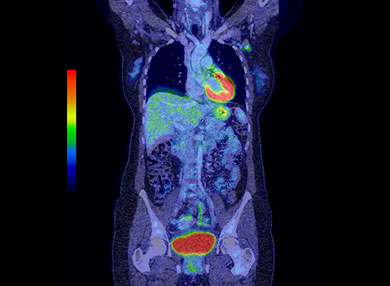
The PET scanner visualises the uptake of this tracer to create an image of the tissue and organs in the body. It can provide valuable information for the diagnosis and treatment of a variety of diseases like cancer, brain or heart disease. It’s also used to investigate epilepsy, Alzheimer’s disease, brain disorders, central nervous system problems, and heart disease.
PET scans are more sensitive in detecting cancer and other diseases than other imaging modalities. PET scanners have a built-in diagnostic CT so that the PET images can be fused (overlaid) perfectly one on the other. These fused images incorporate the sensitivity of a PET scan with the superior anatomy of a diagnostic CT.
By using a combined PET and CT scanner, MMI is able to provide high-resolution CT scans at the same time as a PET scan, saving you time and providing the most useful anatomical and functional information to guide treatment.
The radioactive injection is given directly into your bloodstream via your vein. Different types of radioactive tracer target different types of disease. The PET scanner then detects the tracer and builds a digital image allowing your doctor see what’s going on in your tissues or organs.
The most commonly used form of radioactive tracer is radioactive glucose called FDG. Cancer cells absorb more glucose as compared to the normal cells around it to sustain its abnormal growth rate. The PET scan detects that there is more radiation coming from that area as opposed to the normal healthy cells.
Aside from being able to measure blood flow and oxygen intake, this procedure can check how your body uses sugar.
A PET scan is usually an outpatient procedure. It requires you to fast six hours before the scan.
In preparation for your scan, ensure that you:
- fast (have nothing to eat) for six hours prior to your scan
- if you are diabetic, please download the patient information sheets for more information on preparation
- refrain from any form of exercise for three days prior to your scan
- understand that a PET scan may take up to three hours to complete
- advise staff if you are pregnant or breastfeeding; it is not advisable to breastfeed for 24 hours after the procedure.
Some additional tips to remember:
- If you’ve been taking any medications or vitamins, make sure to tell your doctor about it.
- If you have diabetes, you may eat a light breakfast (e.g. toast and tea/coffee) four hours prior and bring your medication. Your blood glucose reading must be under 10mmol/L for the PET scan to go ahead. Consult your doctor if your blood sugar is above this level with medication.
- If you have a heart condition, don’t take caffeine one day before the test.
- If you are breastfeeding it is best that you express enough milk to be used for 24 hours or use formula for 24 hours after the PET scan.
- Wear comfortable clothes. You may also be asked to wear the hospital gown instead during the procedure. Please remove body jewellery or dentures.
Once the radiotracer is injected, it usually takes up to one hour to course through your body and be absorbed into the organ or tissue to be checked. The scan takes around 30 minutes to an hour to complete.
The physician and radiologist then review the images and write a report, which is sent to your doctor in one to two days.
Introducing Omni Legend
We continually invest in state-of-the-art technology with the aim of providing the best patient experience and the latest imaging technology for specialists.
The new digital Omni Legend PET/CT delivers the highest system sensitivity in its class, and combined with its Precision Deep Learning AI technology, will deliver market-leading image quality, whilst allowing us to deliver significantly lower radiation doses and reduce scanning time.
The scanner provides the highest-resolution images currently available with a lower dose of radiation and offers numerous benefits for specialists.
- Digital PET/CT scanner with a large, 32cm field of view that will significantly reduce scan time, which is especially beneficial for paediatric scans.
- Digital detector produces the highest resolution images in the market.
- High sensitivity digital detection allows us to reduce the radiation dose AND the scan time without compromising on quality.
- Coupled with the digital PET detector is the low-dose CT which enables superior PET/CT image fusion and diagnostic reporting excellence.
- Reporting is provided to specialists within 24 hours.
Patient comfort is important to us. Omni Legend has an array of new features to make the scan as relaxing as possible for patients.
- The new technology developed for the Omni Legend creates PET scans that are at least 50% faster than the previous generation scans, typically under 12 minutes.
- The system features LED ambient lighting to help create a calming mood as well as a graphic pattern on the upper area of the bore.
- MMI bulk bill all Medicare eligible PET/CT studies.